
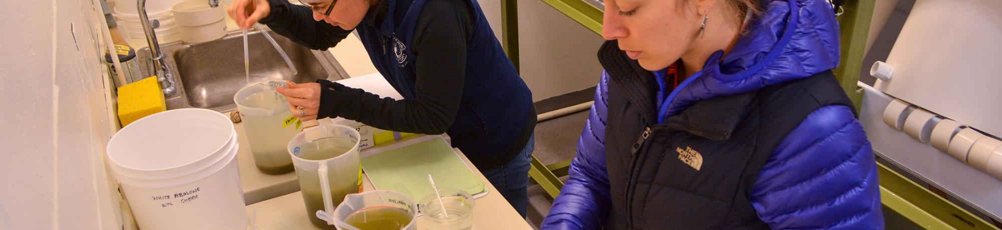
 BML researchers (the Cherr Laboratory) are working to addresses molecules and physiological mechanisms involved in fertilization and early development. This research has focused on fertilization biology using plants, invertebrates, lower vertebrates, and mammals. While significant portions of this research are directly related to how environmental parameters impact successful fertilization and development, much of this body of work focuses on developing a basic understanding of mechanisms associated with sperm-egg interactions and the novel or the conserved aspects of the cell biology.
An example of focus is the study of the mechanisms of sperm motility initiation in Pacific herring from the San Francisco Bay estuary and the egg-derived ligand that signals the intracellular ionic changes and subsequent motility initiation. These sperm have evolved a unique mode of motility initiation in that they surprisingly remain immotile in the environment for hours until they contact an egg, at which time an extracellular glycoprotein stimulates an ionic cascade resulting in flagellar motion. These ionic events are dependent on lowered salinity (in particular Na+) that is typical in the winter months in the SF estuary. Research in the Cherr Laboratory showed that herring sperm motility initiation was due to activation of a reverse Na+/Ca++ exchanger (Proceedings of the National Academy of Sciences USA).
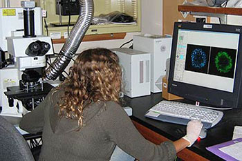
More recently with colleagues from Japan, the Cherr Lab published the model of sperm-egg interaction in Pacific herring that resolves initial mechanistic differences observed between the BML group and those of colleagues from Japan. For much of this physiological research, the BML Fluorescence Imaging Facility has been used. This facility is open to students, postdoctoral scholars, and researchers at BML, as well as those from UC Davis departments. The facility was equipped in part from a multi-user NSF major equipment proposal (Cherr, PI) for our scanning laser confocal microscope (with seawater immersion objectives and temperature controlled stage), while other NSF support was used for high-speed fluorescence imaging systems including an upright fixed stage microscope with microinjection capabilities, a stereo fluorescence system designed for computer controlled high content screening, a fluorometer with live cell capabilities, etc.